Home

hernes ülikond ma olen janune green marker mitochondria facs Ümardage allapoole Purustatud esialgne

Using flow cytometry for mitochondrial assays - ScienceDirect

Parkinson's Disease-Related Proteins PINK1 and Parkin Repress Mitochondrial  Antigen Presentation: Cell
Parkinson's Disease-Related Proteins PINK1 and Parkin Repress Mitochondrial Antigen Presentation: Cell
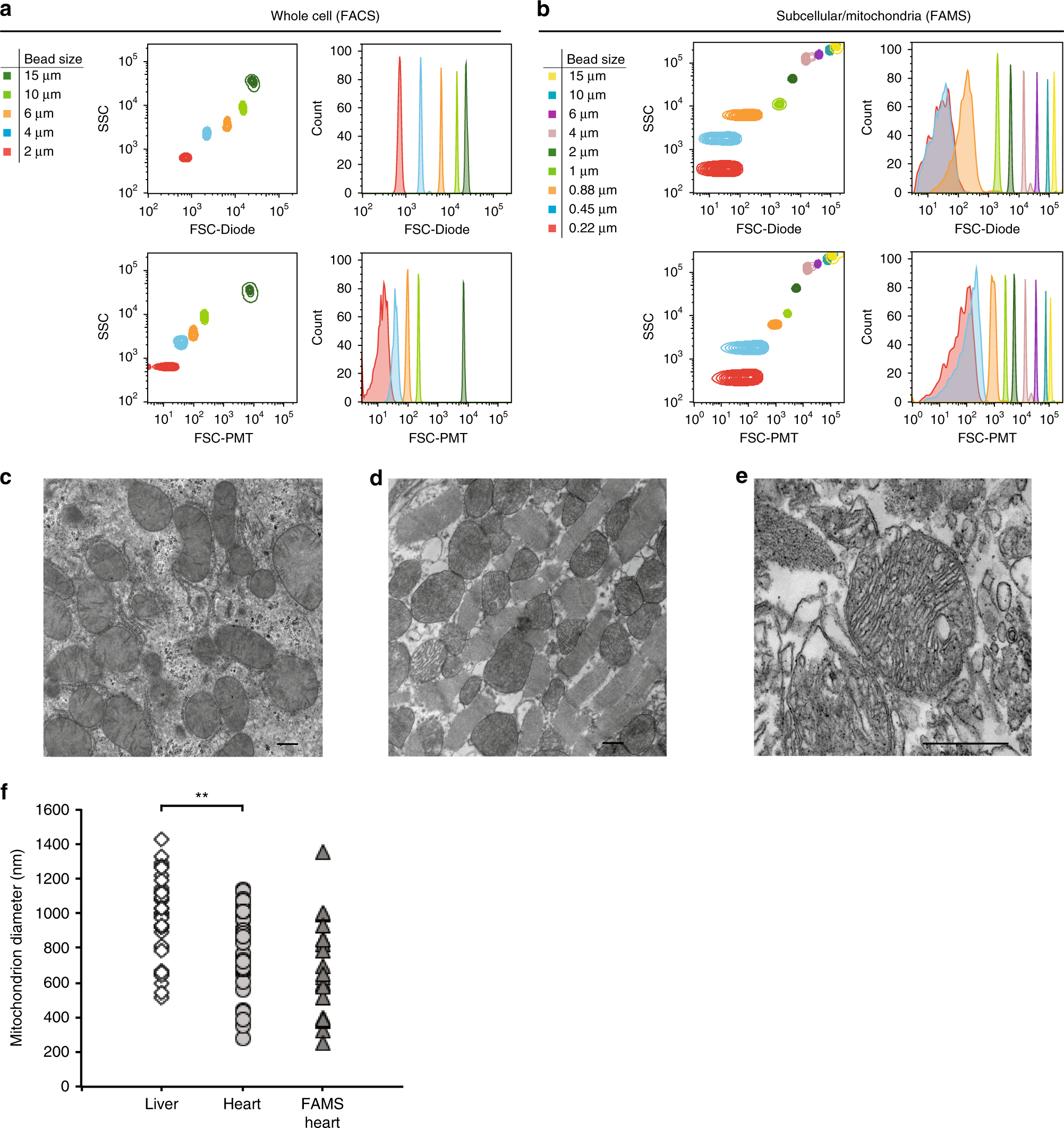
A nanoscale, multi-parametric flow cytometry-based platform to study  mitochondrial heterogeneity and mitochondrial DNA dynamics | Communications  Biology
A nanoscale, multi-parametric flow cytometry-based platform to study mitochondrial heterogeneity and mitochondrial DNA dynamics | Communications Biology

Flow cytometry analysis of MitoTracker Green FM mitochondrial staining... |  Download Scientific Diagram
Flow cytometry analysis of MitoTracker Green FM mitochondrial staining... | Download Scientific Diagram
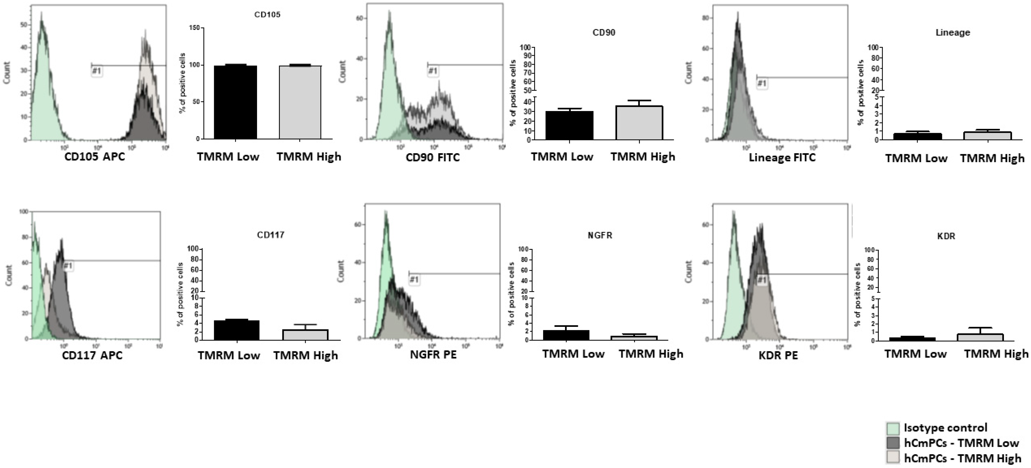
IJMS | Free Full-Text | Differences in Mitochondrial Membrane Potential  Identify Distinct Populations of Human Cardiac Mesenchymal Progenitor Cells
IJMS | Free Full-Text | Differences in Mitochondrial Membrane Potential Identify Distinct Populations of Human Cardiac Mesenchymal Progenitor Cells

Mitochondrial C1qbp promotes differentiation of effector CD8+ T cells via  metabolic-epigenetic reprogramming | Science Advances
Mitochondrial C1qbp promotes differentiation of effector CD8+ T cells via metabolic-epigenetic reprogramming | Science Advances

Cellular Allometry of Mitochondrial Functionality Establishes the Optimal  Cell Size - ScienceDirect
Cellular Allometry of Mitochondrial Functionality Establishes the Optimal Cell Size - ScienceDirect

Cytometric assessment of mitochondria using fluorescent probes -  Cottet‐Rousselle - 2011 - Cytometry Part A - Wiley Online Library
Cytometric assessment of mitochondria using fluorescent probes - Cottet‐Rousselle - 2011 - Cytometry Part A - Wiley Online Library
NIR Mitochondrial Membrane Potential Assay Kit (Flow Cytometry) (ab112149)

JC-10 Mitochondrial Membrane Potential Assay Kit (Flow Cytometry) (ab112133)

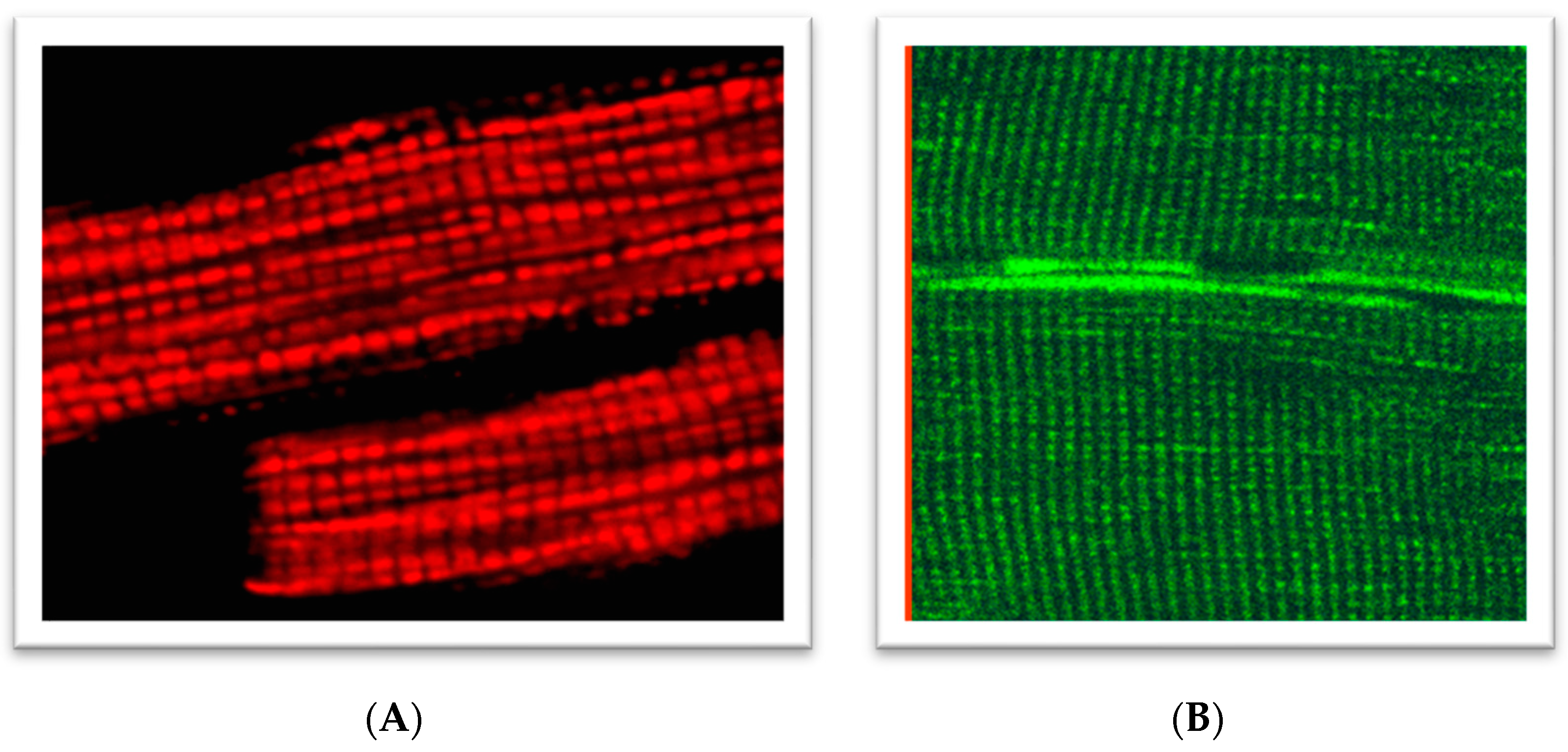
IJMS | Free Full-Text | Analysis of Mitochondrial Function, Structure, and  Intracellular Organization In Situ in Cardiomyocytes and Skeletal Muscles
IJMS | Free Full-Text | Analysis of Mitochondrial Function, Structure, and Intracellular Organization In Situ in Cardiomyocytes and Skeletal Muscles
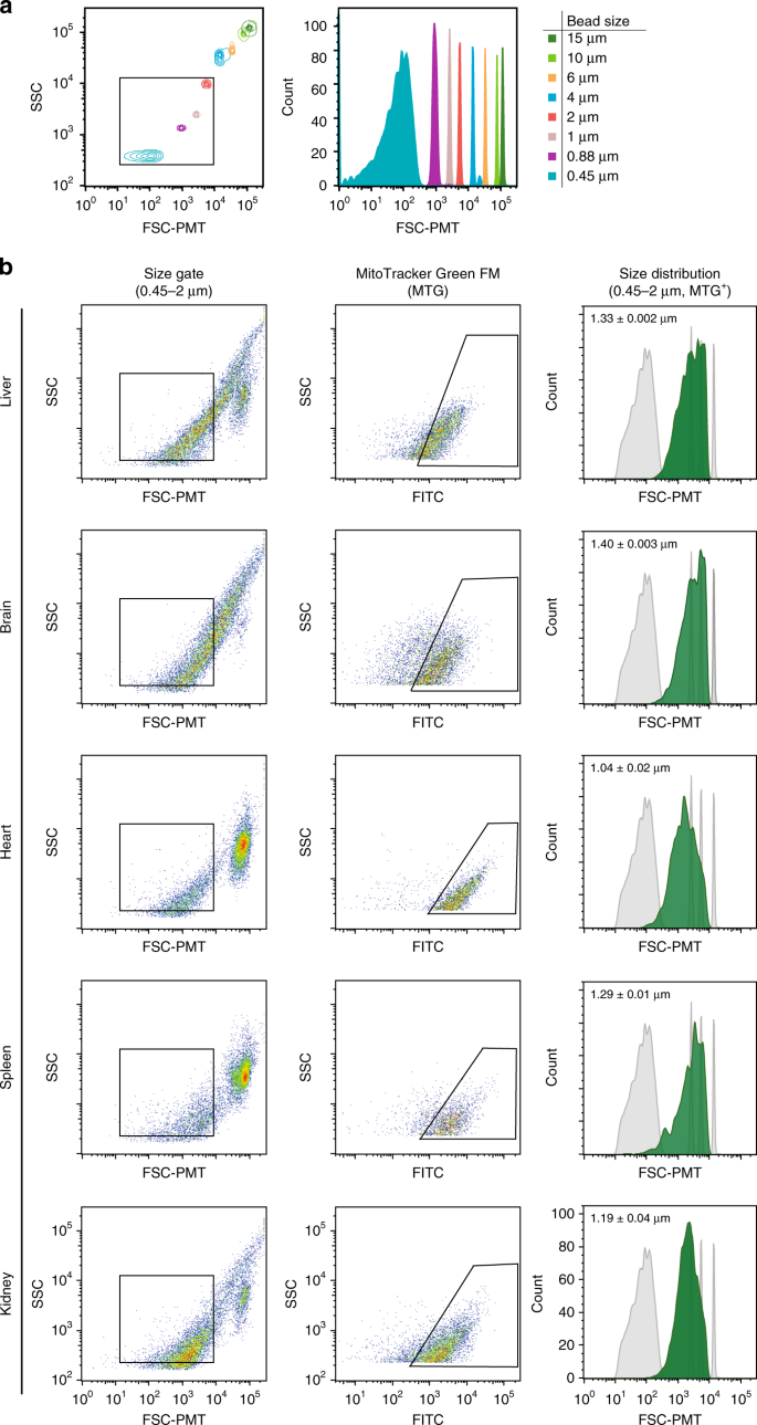
A nanoscale, multi-parametric flow cytometry-based platform to study  mitochondrial heterogeneity and mitochondrial DNA dynamics | Communications  Biology
A nanoscale, multi-parametric flow cytometry-based platform to study mitochondrial heterogeneity and mitochondrial DNA dynamics | Communications Biology
Using flow cytometry for mitochondrial assays - ScienceDirect

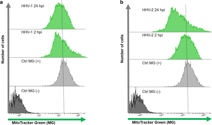
Disturbances of mitochondrial dynamics in cultured neurons infected with  human herpesvirus type 1 and type 2 | SpringerLink
Disturbances of mitochondrial dynamics in cultured neurons infected with human herpesvirus type 1 and type 2 | SpringerLink
Counting Single Cells Using Flow Cytometry

MitoView™ Mitochondrial Dyes - Biotium

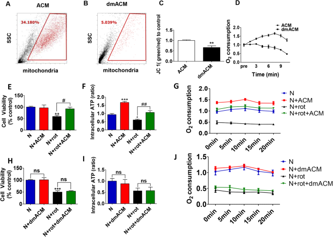
Human iPSCs derived astrocytes rescue rotenone-induced mitochondrial  dysfunction and dopaminergic neurodegeneration in vitro by donating  functional mitochondria | Translational Neurodegeneration | Full Text
Human iPSCs derived astrocytes rescue rotenone-induced mitochondrial dysfunction and dopaminergic neurodegeneration in vitro by donating functional mitochondria | Translational Neurodegeneration | Full Text
Deciphering The Complex Roles of Mitochondria | FlowMetric

Mitochondrial Markers: Novus Biologicals

MitoTracker™ Green FM - Special Packaging

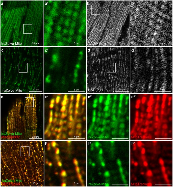
Mitochondrial imaging in live or fixed tissues using a luminescent iridium  complex | Scientific Reports
Mitochondrial imaging in live or fixed tissues using a luminescent iridium complex | Scientific Reports

How to Use Flow Cytometry to Measure Apoptosis: Part One | Bio-Rad

Characterization and analysis of mitochondrial subpopulations by ΔΨ m... |  Download Scientific Diagram
Characterization and analysis of mitochondrial subpopulations by ΔΨ m... | Download Scientific Diagram

Comparison of JC‐1 and MitoTracker probes for mitochondrial viability  assessment in stored canine platelet concentrates: A flow cytometry study -  Marcondes - 2019 - Cytometry Part A - Wiley Online Library
Comparison of JC‐1 and MitoTracker probes for mitochondrial viability assessment in stored canine platelet concentrates: A flow cytometry study - Marcondes - 2019 - Cytometry Part A - Wiley Online Library

Autophagy and Mitochondria - Flow Cytometry Core Facility

MitoView™ Mitochondrial Dyes - Biotium

Deciphering The Complex Roles of Mitochondria | FlowMetric